MEMBRANE-BASED COUNTING OF PARTICULATE
A circular membrane, fixed on a slide, is submitted for analysis according to the USP 788 method.
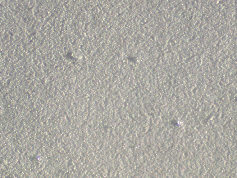
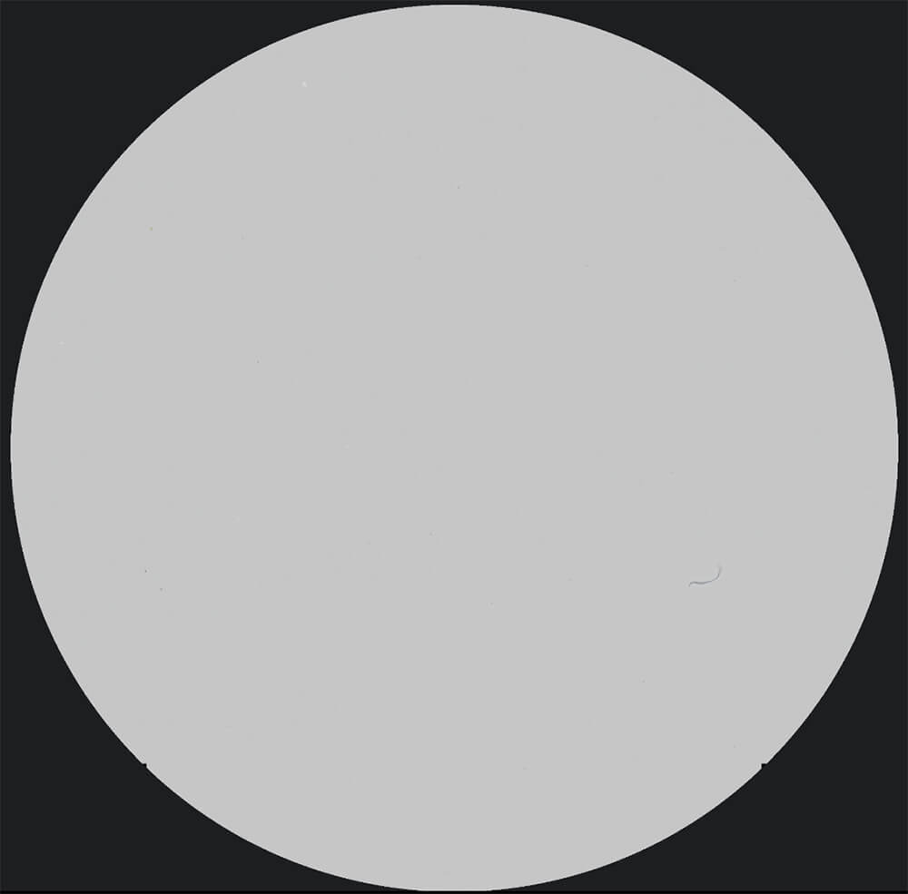
Figure 1. Original image at 100X (1.27 µm/pixels).
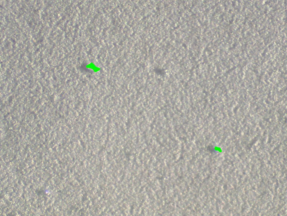
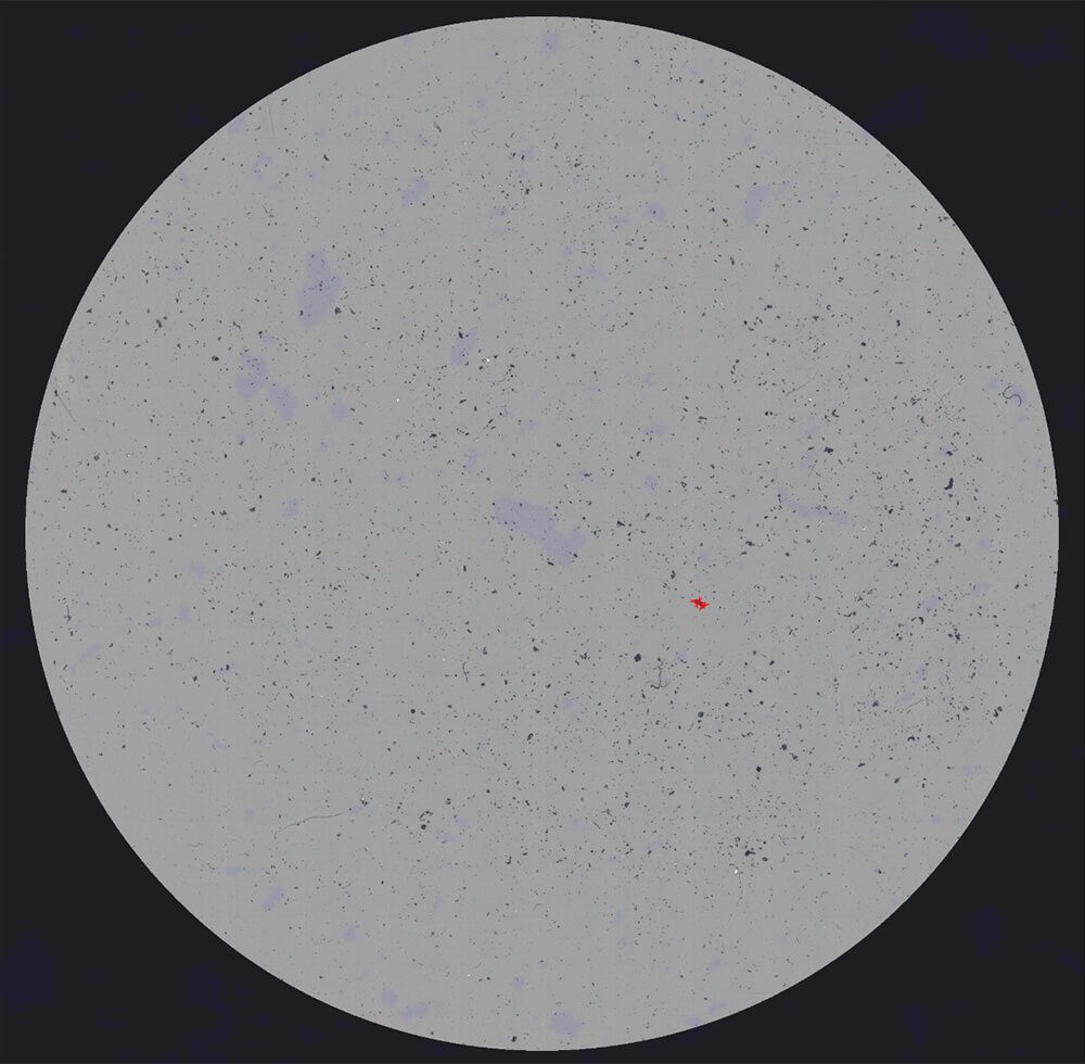
Figure 2. Binarized view of final detected particulates as measured.

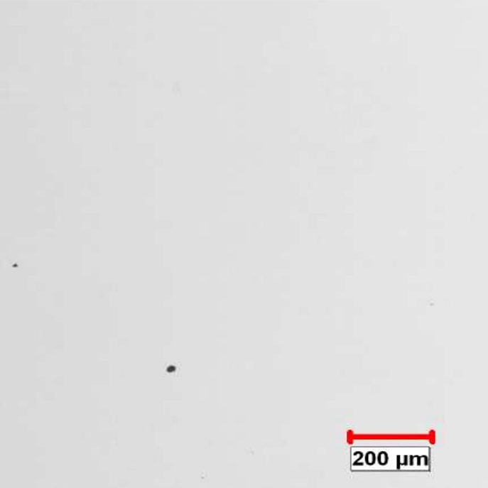
Figure 3. Original image at 100X.
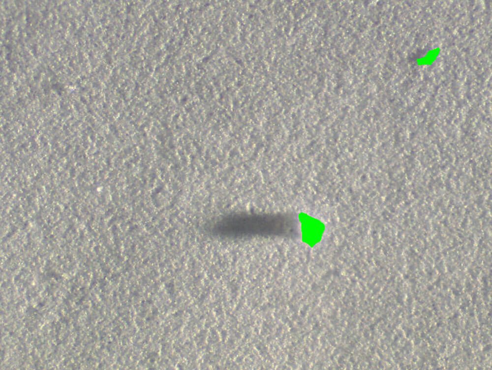
Figure 4. Binarized view of final detected particulates.
PURPOSE
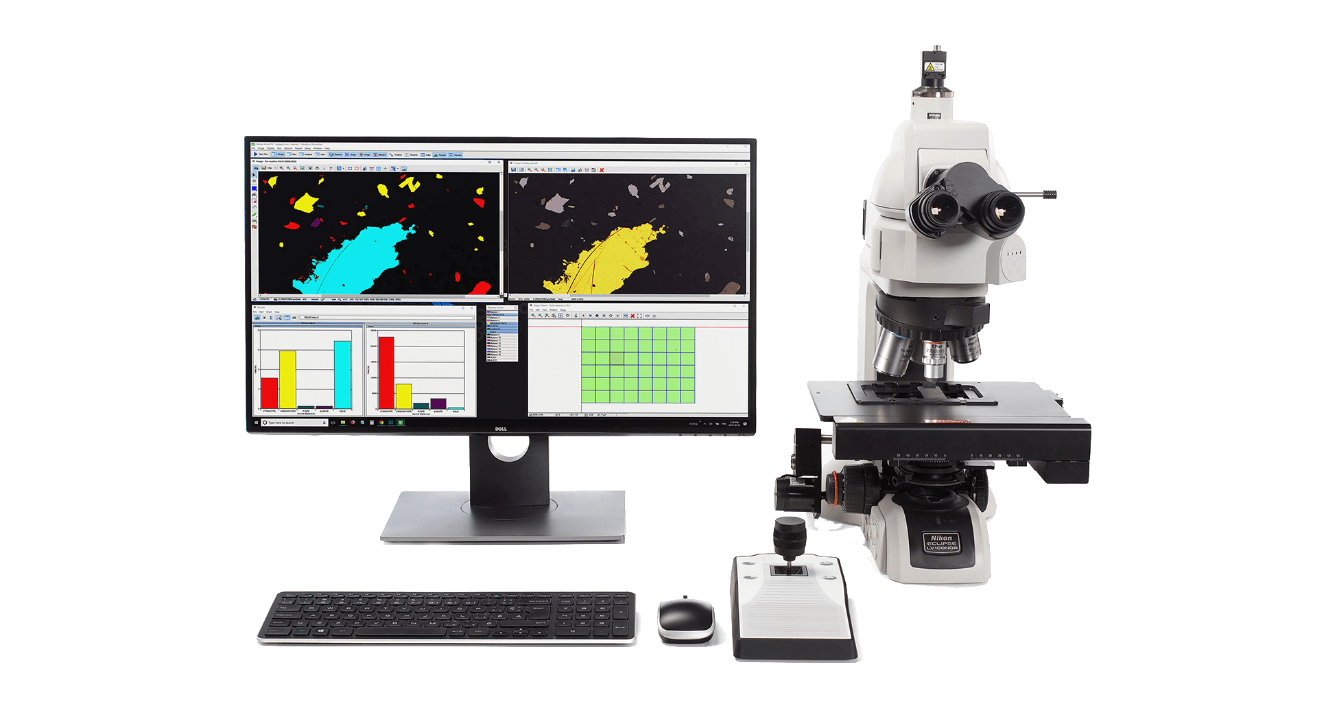
Demonstrate the ability of the Clemex Vision image analysis system can automatically discriminate and measure the particulates deposited on the membrane. The methods and operations used are discussed in the report linked at the bottom of this page (click the Download PDF link below).
RESULTS
Oblique illumination is used in this analysis. The light is projected on the surface at a certain angle to reveal features with higher contrast compared to normal brightfield illumination.
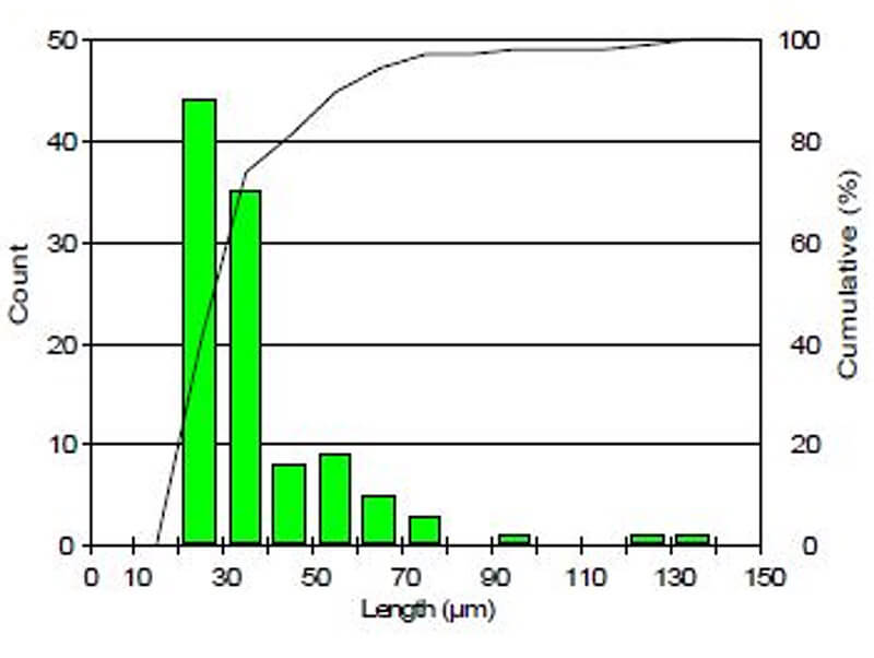
Length measurement is performed on the particulates greater than 25 microns only. A Guard Frame is used to avoid measuring only a part of those features that are sectioned by the field of view. Results were cumulated for automated statistics and graph generation. Final results can be printed directly from Clemex Vision. Raw data can be exported in Excel format.