
Authors Aurélie Chopard, Philippe Marion, Jean-Jacques Royer, Raymond Taza, Hassan Bouzahzah, Mostafa Benzaazoua
Abstract
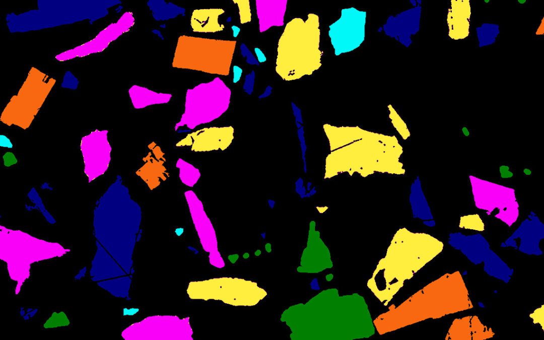
The mining industry needs effective techniques to meet the future challenges of resources extraction. As the deposits become more and more complex, a very good knowledge of an orebody is necessary. Mineralogical characterization is an essential contribution to improve the knowledge on the ore and wastes for a given mining project. It could bring major advances in ore extraction, mineral processing, and integrated waste management. However, mineralogical analyses can be very tedious, when done manually. Consequently, automated mineralogy was developed during the last three decades to improve the rapidity of mineralogical characterization, so that mineralogical information can be routinely obtained. Nowadays, the systems commonly used are based on expensive equipment including scanning electron microscopes (SEM) with energy dispersive X-ray analyzers (EDX). Optical Microscopy (OM) is neglected, although this route can provide reliable and quick results, yet cheaper. In this study, the possibility of using optical microscopy in reflected light mode to automatically characterize opaque minerals is explored. The identification and quantification of six common sulfides from polymetallic ores (arsenopyrite, chalcopyrite, galena, pyrite, pyrrhotite, and sphalerite) were automatically accomplished on a polished section by optical microscopy. Six spectral images were acquired for multispectral image analysis. Five of them were acquired under a white light source, equipped with four different excitation filters (436 nm, 480 nm, 605 nm, and 650 nm). The sixth image was acquired under an UV-light source at 365 nm, after modifying the optical pathway to detect the reflectance of the minerals in the UV-spectrum without changing the acquiring camera. Two image analysis software solutions were then tested to automatically classify and quantify the six sulfide minerals. The classification was systematically done on the acquired multispectral images by grey thresholding with the Clemex Vision PE® software. The GOCAD® software used principal component analysis (PCA) analysis and a supervised K-means clustering method to classify the minerals. Then, the obtained results were compared to the SEM-EDS quantification, considered here as the reference. The differences between the computed surface areas were mainly due to the artifacts of the polished sections preparation. The two software solutions present promising results and could be fully exploited to proceed to other mineralogical analyses, such as mineral liberation, mineral associations, textures identification, and particle size distribution. This study is an initial step towards differentiate and identify sulfides via an optical route arguably reliable and cheaper to characterize mine products.
Purchase full article from ScienceDirect