
Cell Sizing in Extruded Foam
Four pieces of extruded foam were submitted for analysis. Two smaller pieces were pre-cut and used as examples for the current analysis.
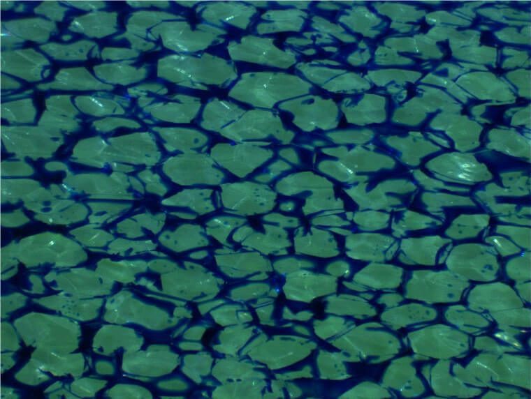
Figure 1. Original image of side A (25x, 5.08 µm/pixel). Cell boundaries are highlighted by blue color.
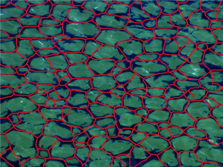
Figure 2. Outlines of cells as measured in red bitplane. Binary operations can be applied on the outlines to convert walls into cells and then perform measurements.
PURPOSE
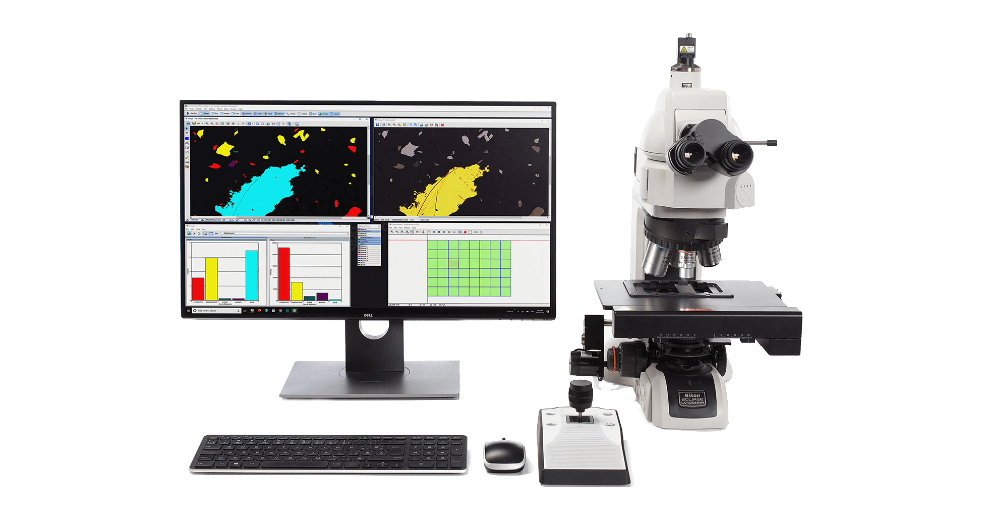
Demonstrate the ability of Clemex Vision image analysis system can distinguish the cells and perform shape, size and oriented size measurements. The methods and operations used are discussed in the report linked at the bottom of this page (click the Download PDF link below).
RESULTS
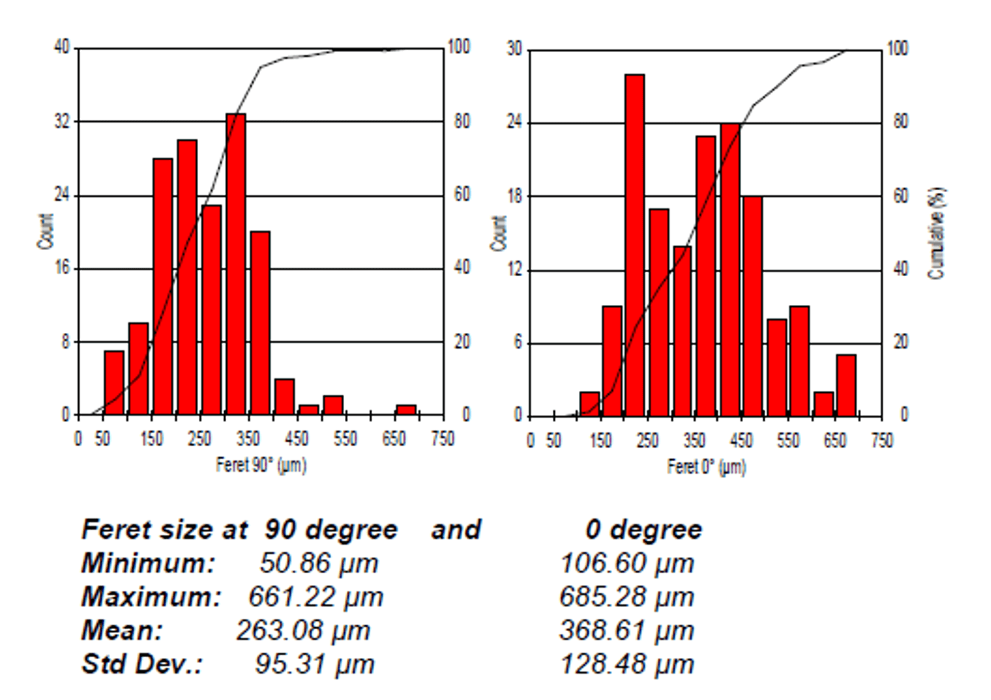
Area, Length, Feret Average, Feret 0°, Feret 45°, Feret 90°, Feret 135°, Circular Diameter, Aspect Ratio and Orientation measurements are performed on each feature. Anisotropy is calculated for each field. Automated statistics and graph are generated and would be cumulated if analyzing several images (samples). Final results can be printed directly from Clemex Vision. Raw data are linked to their respective objects for validation purpose. Raw data can also be exported in Excel format.